
ANALYSIS OF THE MECHANISMS OF ACTION OF ULTRASOUND ON BIOLOGY CELLS
Sultanova G.G
Institute of Botany, Azerbaijan National Academy of Sciences, PhD(Biology)
Associate professor,Leading Researcher,Baku 40,Badamdart Highway,
Baku AZ1004
АНАЛИЗ МЕХАНИЗОВ ДЕЙСТВИЯ УЛЬТРАЗВУКА НА БИОЛОГИЧЕСКИЕ КЛЕТКИ
Султанова Г.Г.
Институт ботаники Национальной Академии Наук Азербайджана,
ведущий научный сотрудник,кандидат биологических наук,доцент
ABSTRACT
The analysis of our own and foreign researches about mechanism of ultrasound (US) action on biological objects is reflected in this article. This researches comprises last 50 years. It’s shown that the biological effects are induced by US action, closely connected with the cavitation and depends of different parameters such frequency, US intensity, compound’s concentration , temperature, pressing and etc. Lysis of cells is under the action of ultrasonic waves is connected probably with the cavitation that is created acoustic microstreams and shearing voltage. It is shown that persistent action of US creates more sufficient changes than impulse one. In connection with the broad application of US in medical practice (physiotherapy, ophthalmology, surgery, internal diseases, microbiology)and food industry it is very important to study the ultrasonic waves action’s mechanism on different objects.
АННОТАЦИЯ
В статье отражен анализ исследований механизма воздействия ультразвука (УЗ) на биологические объекты. Это исследование за последние 50 лет. Показано, что биологические эффекты вызваны действием УЗ, тесно связаны с кавитацией и зависят от различных параметров, таких как частота, интенсивность УЗ, концентрации соединения, температуры, давления и т. Д. Лизис клеток под действием ультразвуковых волн связан, вероятно с кавитацией, которая создается акустическими микропотоками и напряжением сдвига. Показано, что непрерывный УЗ создают более значимые изменения, чем импульсные. В связи с широким применением УЗ в медицинской практике (физиотерапия, офтальмология, хирургия, внутренние болезни, микробиология) и пищевой промышленности очень важно изучение механизмов действия ультразвуковых волн на различные объекты.
Keywords: US-action, cavitation, acoustic microstreams , hemolysis, lysis of cells
Ключевые слова: УЗ-действие, кавитация, акустические микропотоки, гемолиз, лизис клеток
Introduction
Ultrasonic waves of different power are used in the biological researches as physical factor with the selective action on biological objects. The results of experimental data by US action on biomacromolecules, bacteria, viruses, plant and animal tissues are reflected in monography and in papers of some authors – Miller25-27,O’Brien33,Nyborg1,23,24. A lot of experimental data shows that US has specific and selective action on structure of biomacromolecules, cells and cellular membranes. Ultrasonic energy is used efficiently in medicine and microbiological industry. Some different effects are noticed at the US action on cells and cellular suspension:
-heating of US medium (temperature effect);
-chemical damages that are induced by free radicals action in the cavitation process5 and their transformation products;
-mechanic disruptions that are induced by impact waves and acoustic streamings (mechanical factor).
The persistent US action on biological structures gives more effect than impulse one. Let’s see the influence of US field in details on cells in frames of above-mentioned factors. Heat-formation in cellular suspensions takes place in the results of transformation of acoustic energy to thermal as a result of US absorption. The degree of ultrasonic energy depends of medium viscosity, density and of periodic pressing of medium that creates adiabatic increasing of its temperature. Thermal effects may have the particular role in cell’s life. Morphological pictures of cells changes are not similar by the action of heat and US. The border of thermal damaging of tissues less depends of their initial temperature, radiation regime and US frequency. Morphological changes aren’t watched even after 8-hour irradiation if tissues temperature in the US field isn’t more 42-43oC19.Effect of ultrasonic waves action on biological molecules and cells may be connect with the chemical reactions in the US field: 1) process in the gaseous medium; 2) on the border liquid-gas; 3) in the liquid phase in the result of interaction between active compounds of water sonolysis that are diffused in the collapse of cavitation bubbles2,3,5.
Parameters that characterize the chemical reactions in the ultrasonic field depend from different factors: frequency and intensity of acoustic vibrations, temperature and pressure, nature and concentration of dissolved gases.
Both chemical and mechanical US actions take place in the cases when intensity of US vibrations is more than cavitation border’s value. If low-size molecules (monomers, t-RNA, low-size proteins and peptides) are under the US influence the chemical action is over in its value. Mechanical forces play the main role if particles have a large molecular weight (DNA, DNP of viral particles)12. The dependence between effects and intensity of US 8 and time of its action is shown on the fig.1.
The absence of dependence between bactericidal US action on microorganisms suspension and nature of saturated gas gives conclusion that chemical products aren’t significant for cells disruption. Degree of cell disruption isn’t connect with the number of free radicals in the medium under US. Also the absence of dependence between cells disruption and generating free radicals shows that disruptive effects of US on cells defines probably by mechanical factors but not chemical ones 3,8.
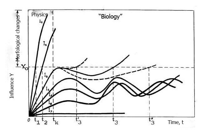
Fig.1. The dependence between biological effects and intensity of US (I1 — >5 Vt/sm2; I2 – 5-1 Vt/sm2; 1,0-0,4 Vt/sm2)
Some authors8,9supposed that prevailing mechanism of cells disruption may be “puncture” of cellular envelope as result of cavitation bubble’s collapse near the cell. The emission of sonic wave is attended by loss of energy that is compensated of acoustic streams. Research of mechanism of US action on bacterial cells by high-frequency filming shows that cells disruption takes place as a result of vortical movement of medium under US action (low-frequency generator, 85 KHertz) 18,20 as simple second search of sound creating fluctuation of cellular membrane.
Acoustic streams may appear near the gaseous bubbles in the US medium that act on the membranes. In the data of Roy 4 amplitude of stabilized oscillating bubble is 30 mk. Microstreams induced by vibratory gaseous bubbles leads the death of microorganisms already in a few seconds of ultrasonic waves action with the amplitude pressure of 0, 1 atm. Some authors40 supposed that cellular suspension under US action create small microstreams near the cell membranes and they may change cell’s structure (fig.2).
There are different changes in the researched object in the result of mechanical action of US cavitation. These changes are in dependence of US-parameters, irradiation conditions and cell’s state. Application of US based on US capability to create instantaneous disruption of bacteria, animal, plant cell and within cellular structures20.
It’s shown that only ultrasonic waves of definite intensity have disruptive action on cells. The border of cells disruption is defined by their nature, concentration and ultrasound conditions. Acoustic streams in suspension are appearing on pre-cavitation regime (0,05Vt/sm) and are capable only to “wash” macromolecules from surface of cell membranes. US intensity increases till values more than border of cavitation and forms the pulsing of gaseous bubbles in the medium, creating microstreams with the speed gradients 104 c -1 7.
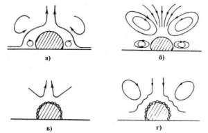
Fig. 2. The diagram of appearance of acousting streams.
Mechanical disruptions may be conditioned by shearing voltage in appearance of acoustic streams with the high speed gradients 104-105с-1 that will reach of 3-5х103 dyne/sm2 14,28.
The presence of gaseous microbubbles of resonance sizes in the medium increases the efficiency of US action in considerable extent.
There are gaseous bubbles in the regime of US action (intensity J= 1 Vt/sm2 and frequency f=1 M Hertz) on cells, within cells or near the plasmatic membranes. These bubbles may leads to vibration in the cellular membranes in microstream. Thrombocytes begin to aggregate under the US action with the frequency of 1M Hertz and intensity 32 MVt/sm2 if the shearing voltage will reach 50 dyne/sm2 23,24.
Leucocytes disruption in the field of stabile pulsating gaseous bubbles which frequency (about 20 KHertz) begins with the vibration amplitude increasing (its value is till 3). Process of haemoglobin extraction from erythrocytes at the 15-20min of US treatment conditioned of the creation of a large hydrodynamic forces near the vibrated bubble 8,28.The mechanism of disruption usually has also mechanical nature at the high-frequency US action (about1 MHertz ) on cell suspension.The border of US intensity that cases cells death is depended both of US frequency and type of cells 3,8.
There are shown that prevalent role in the modifying US action on cells have mechanical forces that are appearing in bubbles collapse-impact waves and hydrodynamic streams and microstreams.
When the frequency and intensity of US vibrations are varying and also the duration of US treatment it may be possible to control of these processes. US doesn’t only disrupt the cells but it may stimulate and increase of cells vitality that were in poor physiological state before, inhibition of cells division, alienation of cells death, increasing of cell membranes permeability, conformational changes in DNA molecule and others.
It’s mentioned that continuous US creates more sufficient changes in cells structure that impulse one10,22. In the researches of US biological action usually the main parameters of it are intensity and total time of action 1-3,8.
There are changes in diffusion processes, intracellular viscosity, membrane conductance in the intensity 0,1-1,0 Vt/sm2 and also there is the process of sonoluminiscence and biologically active substances and radicals are creating. Efficiency of US influence on cells and cellular structures also depends of microorganism’s type, medium content, cells concentration, their morphological features, sizes, forms and functional state10,16.
Sensitivity of different cells to US action varies very much. Amoeba’s cells exist in the intensive radiation: there are 50% of vitality individuals in suspension (18) after 10 min. of US treatment (200 Vt/sm2 (1MHertz) and also cells in suspension begin to disrupt at the 0,7 Vt/sm2 (0,75 МHertz, 1min.)31.
Braginskaya3has shown that there isn’t correlation between increasing of dead cells number and decreasing of vitality cells number because some cells under US
treatment are deformed( they aren’t viable but cann’t characterized as dead).
Some appreciable changes of physiological state are before the disruption of cells structure and if mechanical resistance of cells changes in wide limits (0,1-1,0 Vt/sm2 1-104 s. at the 0,5-2 МHertz) in dependence of cells type, the border (threshold) of physiological changes also (0,1-0,5 Vt/sm2 10-100 s. at the 0,5-2 МГц) isn’t change. First of all cells resistance to US defines by the structure of their cellular envelope that is most under the influence of factors in US field. It’s shown that US (0,88MHertz, 0,6 Vt/sm2) in certain conditions sufficiently (on 60-100%) increases conductivity of bilayer lipid membranes and also their permeability for anion of borate tetraphenyl 7.
It’s also discovered that focused US increases involving of channel-former antibiotic nystatin into matrix of phospholipid membranes (on 10%) that may be conditioned by changes in matrix mechanical properties and damages in the neighboring diffusion layers 18.
US effects not only on the processes of cell’s vitality but also on structure and function of some cellular organelles. There are some damages in lysosomes membranes under the US action (2,5 Vt/sm2, 1 МHertz, 1 min.) and lysis of cells in the rats liver 30. We were watching the parallel effect in the action of low-frequency US (20KHertz) 10. US in certain conditions (0,75 МHertz, 2 Vt/sm2) may disrupt nucleus in cells but doesn’t disrupt integral cytoplasmatic membranes 32,33. These disruptions aren’t conditioned of cavitation and microstreams and probably are explained by creation of resonance waves on the surface of nuclear membranes.
The created changes are repaired during 100 hours if US action isn’t lethal for the cell and only mitochondrions need more time for repairing of their structure and function 30.
US action on cell isn’t limited of influence on surface structures. There are power microstreams in the US field that are mixing the cell’s content and changing the place of cellular organelles 10. The source of these microstreams may be vibration of cytoplasmatic membrane or pulsating gaseous bubble if the distance between source and cell isn’t more than 5х10-2sm 24,26.
We discovered that during the long time there is development of processes post-morphological and post-functional interruptions in the cell after US radiation. For example electrophoretic mobility of erythrocytes decreases after US treatment (0,02-1Vt/sm2; 0,8 МHertz и 0,4 МHertz, 3-180sec.) and repaires in 3-5 min after US switching.
Thrombocytes after US treatment (1 MHertz, 0,2-0,6 Vt/sm2, 5 min.) on the electron microphoto aren’t differ from control ones. They are functioning as intact thrombocytes and form thrombuses but in control and US irradiated cells there are functional and morphological differences after 30 min. of incubation at the 220С 26. The time of recalcification of thrombocytes less changes during the process of US treatment(1 МHertz, 0,065-2 Vt/sm2, 5 min) and decrease in 4-6 hours after irradiation 27.
US-treatment of the cells in suspension or culture depends of US parameters and irradiation conditions and may be the reason both the stimulation and depression of vitality processes. US of biochemical processes in the cells increases biological activity and resistance to environment. The marrow cells were irradiated by US (0,8MHertz, 0,3-0,7 Vt/sm2, 20 min) and injected to control animals; they give the beginning of a large number of colonies on the surface and in the spleen’s parenchyma. These colonies grow faster and colonies differentiation increases in this case27.
US treatment (0,9 и 2,6 МHertz, 10 min, 0,5-1,2 Vt/sm2) less decreases the number of vitality cells in marrow suspension but after some days of their storage at the 40C in US-irradiated probes there are more vitality cells than in control ones.
The time of disruption of a half of marrow cells in suspension after US treatment increases twice – from 5 to 9 days that decrease losses in cells storage that may be use in transplantation 3.
The increasing of US intensity till values more than 1,0-1,5 Vt/sm2 as a rule inhibits the cells biological functions. Continuous US (1МHertz, 0,8-2,6 Vt/сm2, 60 min) inhibits the speed of growth of amniotic human cells
in culture and the threshold of inhibitory action is between 0,8 and 1,7 Vt/sm2 24,34.
Usually animal cells in culture exist good US irradiation except cavitation or heating. Survivor cells as a rule are capable to normal growth and development but colonies created by these cells sometimes reach the sizes of ones that were formed from unirradiated US cells16.
US action also decreases the speed of cells division. Animal cells are more sensitive to US and the speed of cells division decreases at the very low US intensity. So five-minute of US-irradiation with the intensity of 60 МVt/sm2 and with the frequency of 1 MHertz inhibits mitotic index in tissues of rats liver 21.The US irradiation ( 0,1Vt/sm2, 2МHertz, 5 min.) of Erlich adenocarcinoma cells inhibits tumor’s growth that was forming at the reinjection of these cells3
US may increase the processes that are going more slowly in certain conditions 8.US is used for intensification of some processes that are connected` with the fermentation3 and make possible to solve some technological problems.
Conclusion
We use this information and may consider that the main parameter of the process is only duration of US if you choose stationary regime at the disintegration of cells by US action (concentration of cells, US intensity, volume of suspension, distance of immersion of the oscillator head to medium). The inductions time is very important factor in the process of US action; during of this period the speed of destruction is very small and replace the fast one of transformations in the suspension after US treatment. Processes in the US medium in induction period may be essentially for the next transformations in cells in US field.
References
- Nyborg W.L. Biological effects of ultrasound: development of safety guidelines. Part II: general review.(2001) Ultrasound Med Biol. .V27, 3, p.301-33.
- Sultanova G.G. About the mechanism of ultrasiond action in cavitation regim.(O mexanismax deystviya ultrasuka v usloviyax kaitaciyi)(in Russian). Journal of Qafqaz University,Chemistry and Biology,2015 v. 3 № 1, p.9-16,2015
- E. A. Brujan, T. Ikeda, and Y. Matsumoto. Jet formation and shock wave emission during collapse of ultrasound-induced cavitation bubbles and their role in the therapeutic applications of high-intensity focused ultrasound. Physics in Medicine and Biology, 50(20):4797, 2005.
- C. C. Coussios and R. A. Roy. Applications of acoustics and cavitation to noninvasive therapy and drug delivery. Annual Review of Fluid Mechanics, 40:395–420, 2008.
- Margulis М А , Margulis I M ( 2005) Regarding of mechanism of biological action of ionizing radiation (in comparison with ultrasonic caitation). J. of Physical Chemistry 79, 6, 1142-1151( Russian)
- S. Vaezy and V. Zderic. Hemorrhage control using high intensity focused ultrasound. International Journal of Hyperthermia, 23(2):203–211, 2007.
- Sultanova G G, Samedova A A , Zeynalova N M , Abdulova X D ( 2004) The effect of some fiziologically active substances on erythrocyte membrans. XVII Ulusal biology kongresi, CukurovaUniversitesi Adana, 7
- C. R. Hill, J. C. Bamber, and G. R. ter Haar, eds. Physical Principles of Medical Ultrasonics, 2end.. Wiley, 2004. 511pp
- M. Gy¨ongy and C.-C. Coussios. Passive cavitation mapping for localization and tracking of bubble dynamics. The Journal of the Acoustical Society of America – Express Letters (submitted), 2010.
- K. G. Baker, V. J. Robertson, and F. A. Duck. A review of therapeutic ultrasound: Biophysical effects. Physical Therapy, 81:1351–1358, 2001.
- Aradniv M, Doida X, Miller M W, Brayman A A, Meltrer R S.(1996) Temporality of ultrasound induced cell lysis in vitro. Echocardiography, 13, 1,45-56
- Sultanova G G (2017) Biological characteristics of ultrasonic effects of biomacromolecules and cells
- C.C.Cousios, C.H.Farny,G.Ter Haar and R.A.Roy. Role of Acoustic Cavitation in the Delivery and Monitoring of Cancer Treatment by High- Intensity Focused Ultrasound (HIFU) Int. J. Hyperthermia, 2007; 23(2): 105–120.
- Jagannathan S, Berlyn R.Mellein, and S.Mitragotri An Experimental and Theoretical Analysis of Ultrasound-Induced Permeabilization of Cell Membranes, Biophys J. 2003 V 84,№5б, р 3087–3101.
- Larrat B., Pernot M., Aubry J., Dervishi E., Sinkus R., Seilhean et all. MR-guided transcranial brain HIFU in small animal models. Physics in Medicine and Biology, 55:365–388, 2010.
- Champion J V, North P F, Coakley W, Williams E(1981) Shear fragility of human erythrocytes. — Biotechnology 8, 1,23-29.
- Coakley WT., Hampton D, Dunn T (1971) Quantitative relationships between ultrasonic cavitation and effects upon amoeba at 1MHz. J. Acount. Soc. America 50, 62, 1546-1553.
- C. H. Farny, R. G. Holt, and R. A. Roy. The correlation between bubbleenhanced HIFU heating and cavitation. IEEE Transactions on Biomedical Engineering, 57:175–184, 2010
- K. G. Baker, V. J. Robertson, and F. A. Duck. A review of therapeutic ultrasound: Biophysical effects. Physical Therapy, 81:1351–1358, 2001.
- B. Larrat, M. Pernot, J.-F. Aubry, E. Dervishi, R. Sinkus, D. Seilhean, Y. Marie, B. A-L, M. Fink, and M. Tanter. MR-guided transcranial brain HIFU in small animal models. Physics in Medicine and Biology, 55:365–388, 2010.
- Kremkau, F.W., Barnes, R.W. and McGraw, C.P. (1981). Ultrasonic attenuation and propagation speed in normal human brain. J. Acoust. Soc. Am. 70, 29–38. .
- Lautesborn W (2004) General and basic of cavitation. — In books: “Finite amplitude wave effect in fluids”,.
- Miller D L , Nyborg W I , Whatcom C C ( 2012) In vitro clumping of pantalets exposed to low intensity ultrasound. In: Ultrasound in Medicine (Ed. D. White, E. Lyons), N-Y, L, Plenum Press, 545-553.
- Miller D L , Nyborg W I, Whatcom C C ( 2012) Bioeffects of ultrasound “Low intensity” condirions. — In: Ultrasound in Medicine (Ed. White, E. Lyons), N-Y, L, Plenum Press, 571-573.
- Miller M.W, Brayman A A, Sherman T A, Abramowicz J C,Cox C( 2001), Comparative sensitivity of human fetal and adult erythrocytes to hemolysis by pulsed 1 MHz ultrasound, Ultrasound in Med. and Biol.27,3,419-425
- Miller M W , Everbach E C, Cox C, Knapp R R, Brayman A A , Sherman T.A.( 2001) A comparison of the hemolytic potential of optison and albunex in whole human blood in vitro: acoustic pressure, ultrasound frequency, donor and passive cavitation detection considerations Ultrasound in Med. and Biol. 27,709-721.
- Miller M W, Sherman T A, Brayman A A ( 2000) Comparative sensitivity of human and bovine erythrocytes to sonolysis by 1-MHz ultrasound, Ultrasound in Med. and Biol.26 ,8,1317-1326.
- C. H. Farny, R. G. Holt, and R. A. Roy. The correlation between bubbleenhanced HIFU heating and cavitation. IEEE Transactions on Biomedical Engineering, 57:175–184, 2010
- C. C. Coussios and R. A. Roy. Applications of acoustics and cavitation to noninvasive therapy and drug delivery. Annual Review of Fluid Mechanics, 40:395–420, 2008.
- V. A. Salgaonkar, S. Datta, C. K. Holland, and T. D. Mast. Passive cavitation imaging with ultrasound arrays. The Journal of the Acoustical Society of America, 126(6):3071–3083, 2009
- N. McDannold, N. Vykhodtseva, and K. Hynynen. Targeted disruption of the blood-brain barrier with focused ultrasound: association with cavitation activity. Physics in Medicine and Biology, 51(4):793–807, 2006.
- Wibble J H,Galen K P,Wojdila JK.et.al Microbubbles induce renal hemmorage whenexposed to diagnostic ultrasoundin anestized rats Ultrasound in Med. and Biol,2002,28,1535-1546
- W. D. O’Brien. Ultrasound-biophysics mechanisms. Progress in Biophysics and Molecular Biology, 93(1-3):212–255, 2007.
- M.H. Repacholi, Martino Gandolfo, A. Rindi .Ultrasound: Medical Applications, Biological Effects and Hazard Potential, Springer Science & Business Media, 2012 p.386
- J. E. Kennedy. High-intensity focused ultrasound in the treatment of solid tumours. Nature Reviews Cancer, 5(4):321–327, 2005.
- H.G. Zhang, K. Mehta, P. Cohen, and C. Guha. Hyperthermia on immune regulation: a temperature’s story. Cancer Letters, 271:191–204, 2008.